Friday, February 29, 2008

Tracheostomy


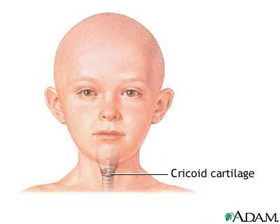
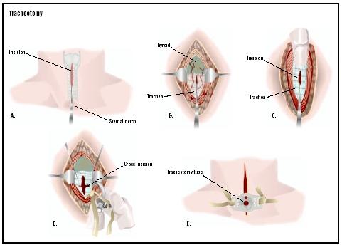
Landmarks for tracheostomy


  1. Cricoid cartilage - It is a circular cartilage below the thyroid cartilage and is the shape of a tyre. It is tne only cartilage in larynx and trachea which forms a complete ring and also it forms the widest ring and the most prominent one.


  2. Suprasternal notch - Make an incision two finger breadth above supra sternal notch.


  3. Strap muscles - these are separated vertically by blunt dissection.


  4. Isthmus of thyroid gland - If possible retract it upwards or if not put two clamps, cut and transfix it.


  5. Tracheal rings - palpate and confirm by aspirating with a syringe.

Thursday, February 28, 2008

Tracheostomy

High tracheostomy
If the opening in trachea is made between 1st and 2nd ring, it is called high tracheostomy. The main indication for a high tracheostomy is carcinoma larynx. As stoma is at a higher level and if it gets involved , it can be removed along with the larynx leaving good amount of trahea for making airway. If it is done in other cases, it can result in tracheal stenosis which is very difficult to deal with.

Wednesday, February 27, 2008

Tracheostomy


Procedure -


Position of the patient - The patient is lying supine with a sand bag below the shoulder so as to extend the neck and make the trachea prominent but in an already respiratory compromised patient, the patient becomes more uncomfortable with this posture.


The neck is painted with povidone iodine solution and sterile sheets placed. The area is infilterated with 2% lignocaine with adrenaline solution in a diamond shape fashion. The area is palpated and landmarks identified. The incision is placed two fingers above the suprasternal notch. Most of the times a transverse incision is given in a skin crease but in emergencies a vertical incision is of great help. The subcutaneous tissue is divided and muscles exposed. Great care is taken not to leave the midline otherwise one can land in complications. The fat which comes in the way can be safely removed to get a better exposure.The strap muscles are neatly separated in the midline with blunt dissection with a small artery forceps. Now the isthmus of thyroid comes in the way. This can be lifted up with a hook or clamped with long artery forceps, cut and transfixed with ligatures.The pretracheal tissue is gently dissected and trachea exposed and identified from tracheal rings. Small amount of local anaesthetic is injected in trachea to decrease the cough reflex after aspiration of air to confirm the position of needle.Now the trachea is incised between 2nd and 3rd tracheal or 3rd and 4th tracheal rings.A small piece of cartilage can be removed to facilitate the entry of tracheostomy tube but in children this is avoided. Now the tracheostomy tube is inserted and secured. The wound is closed with silk or prolene sutures.

Tuesday, February 26, 2008

Tracheostomy VS Intubation

Following are the advantages of intubation and disadvantages of tracheostomy-
  1. It is easier and quicker to perform as compared to tracheostomy.
  2. It less invasive.
  3. No scar formation is there.
  4. Drugs like adrenaline can be given through it.
  5. No bleeding from the surgery site is there.
  6. No complications like pneumothorax or delayed complications like tracheo-oesophageal fistula is ther.
  7. Can be easily removed, no complication of difficult decanulation.

TRACHEOSTOMY VS INTUBATION

Following are the benefits of tracheostomy over endotracheal intubation-

  1. It makes the patient more comfortable and less irratable.
  2. It reduces need for sedation.
  3. It improves ability to maintain oral and bronchial hygiene. It helps in tracheo-bronchial toilet.
  4. It reduces risk of glottic trauma. There can be injury to vocal cords and also intubation granuloma formation with intubation.
  5. Tracheostomy reduces dead space and reduces work of breathing.
  6. It can be kept for prolonged period of time.
  7. It augments process of weaning from ventilatory support.

Sunday, February 24, 2008

TRACHEOSTOMY

Indications of tracheostomy
  1. For felieving upper airway obstruction - this is done in cases of carcinoma larynx and hypopharynx when these are obstructing the airways and also after laryngectomy for maintainance of airways. Also in cases of foreign bodies and diseases compromising airways like dyptheria and retro and para pharyngeal abscesses, ludwig's angina etc
  2. For tracheo-bronchial toilet in cases of long standing illness like CVA
  3. For assisted ventillation
  4. To decrease the dead space
  5. For other surgeries like long surgeries of oro maxillary areas, and in cases where patient is not able to open mouth like TM joint ankylosis or mass obstructing in the oral cavity and pharynx
  6. For treatment of diseases like obstructive sleep apnoea.

Saturday, February 23, 2008

TRACHEOSTOMY


The other tracheostomy tubes are non metallic ones.There are two types of non metallic tubes, one cuffed and other is non cuffed. The advantage of cuffed tubes is that these seal the lumen of trachea due to inflated cuffs and no aspiration of secretions in tracheo-bronchial tree is there and secondly when the patient is on positive pressure ventilation ,no leakage of air takes place. The disadvantage is that the cuff should be deflated at regular intervals otherwise chances of tracheomalacia are there. The non metallic tubes are made of inert material so less foreign body reaction and they can be easily connected to ventilator. As there is no phonation opening, patient is not able to phonate in initial stages.The tube has a radio opaque line which helps in location of the tube on x-rays.

Friday, February 22, 2008

TRACHEOSTOMY


There are different types of tracheostomy tubes available in the market. Basically there are metallic and non metallic tubes. Metallic tubes have different shapes and sizes. Fuller's metallic tube has an inner tube and an outer tube. Inner tube is longer than the outer tube to prevent crusting. It has an opening on its shoulder for phonation. The outer tube is bi- phalanged.The purpose of phalanges is that the tube can be easily inserted by pinching the phalanges. The advantage of this metallic tube is that its insertion is easy. Secondly if inner tube is removed for cleaning purposes, the outer tube remains in the trachea to maintain respiration.Another advantage is phonation. The patient can learn to close the opening of tracheostomy tube with the finger and phonate as the inner tube has an opening for phonation.The disadvantage is that ventilators are difficult to connect with these tubes. Secondly these can cause pressure necrosis of trachea and the tube can itself get erroded and fall in the trachea causing a foreign body in airway.

Thursday, February 21, 2008

CRICOTHYROTOMY


Cricothyrotomy is a life saving procedure. If a patient is having sudden respiratory obstruction and no particular instruments or fascilities are available then this procedure is of great help even in the hands of non medical people. In this, a slit is made in between the thyroid cartilage and cricoid cartilage. Thyroid cartilage is also called Adam's apple. It is V shaped and can be easily palpated in the neck.Cricoid cartilage is a round structure below the Adam's apple and is like a small tyre. The space in between these two cartilages is cricothyroid space and is covered by cricothyroid membrane. It is this space and membrane through which we make an opening in the larynx. With a blade or in emergency with any sharp object like knife after palpating the cricothyroid membrane a slit is made and widened. When no instrument is available the tip of a pen can puncture this space and then it is made patent with the hollow of a pen.Otherwise after widening the slit, a trcheostomy tude or endotracheal tube can be inserted through it and respiration secured.

Wednesday, February 20, 2008

TRACHEOSTOMY


Tracheostomy is a life saving procedure in which a hole is created in the trachea (wind pipe) for respiration in cases of airway obstruction and a trachestomy tube is inserted in the trachea through it which bypasses the upper respiratory tract. In case of emergency due to respiratory tract obstruction,this is the procedure which can save the life of a patient and every health worker whether he is a paramedic or a doctor should know how to perform it in case of emergency.


Another procedure which is quick, easy and used when no special instruments are available is cricothyrotomy or tracheotomy. In this a hole is made between the thyroid cartilage and cricoid cartilage.

Tuesday, February 19, 2008

TYMPANIC MEMBRANE


Tympanic membrane commonly known as drum separates the external ear from the middle ear. It is pearly white in color and is semitranslucent. There are some landmarks on the tympanic membrane which are -

  1. Handle of malleus which slants in posterior direction.
  2. Cone of light which gets reflected in anteroinferior quadrant.
  3. Umbo - the most prominent part of tympanic membrane
  4. Anterior and postrrior malleolar folds - above these the tympanic membrane is called Pars Flaccida or sharpnell's membrane and below these is called pars tensa.
  5. Long process of incus and incudo-stapedial joint.
  6. Anterior process of malleus.

Saturday, February 16, 2008

MYRINGOPLASTY-INDICATIONS


The grafting of drum in case of perforation is necessary due to following reasons-


  1. As the tympanic membrane has a hole in it,the chances of getting water from outside into the middle ear through the perforation are always there while bathing or swimming.This will contaminate the middle ear and cause infection in it and the ear will start discharging pus.So a route of infection persists if the perforation is not closed. The patient has to leave the sport of swimming.Also if the patient has a discharging ear, it becomes a sourse of embarasement for him in public life.

  2. Due to perforation, the patient suffers some degree of hearing loss which depends on the site and size of perforation. If the perforation is small but is in the posteroinferior compartment opposite the round window area, itwill produce more hearing impairment.

Friday, February 15, 2008

MYRINGOPLASTY-COMPLICATIONS


Ther are a few complications of myringoplasty also. If done under general anaesthesia, the complications due to anaesthesia can occur. Then there can be injury to a small nerve which supplies anterior 2/3 of tongue and carries taste sensation to brain. This nerve is chorda tympani and injury to this causes altered taste sensation. Then post operative vertigo can be there which lasts for short time. Infection can occur and this can also lead to graft rejection. Then a small residual perforation can remain after surgery.

Thursday, February 14, 2008

CLOSING SMALL PERFORATION OF TYMPANIC MEMBRANE-II


Another method of closure of tympanic membrane perforation is by fat graft. The edges of the perforation are made raw by removing a thin margin after giving local anaesthesia. After freshenening the margins, the fat is harvested from the ear lobule. A small incision is given on the lateral surface or under surface of lobule of ear and fat taken out of the lobule. This fat is placed in the perforation in such a manner that a part of this remains above and a part below the perforation like a dumbell. The ear canal is packed with gelfoam.

Wednesday, February 13, 2008

CLOSING SMALL PERFORATION OF DRUM


Many a times in our clinical practice, patients come with small tympanic membrane perforation 2-3 mm in size. In these patients surgery is not required and one can close the perforation by other techniques also. If the ear is dry, one can cauterize and raw the edges of the tympanic membrane by applying Trichol acetic acid (TCA). As the outer layer of drum which is epithelium and inner layer that is mucosa meet at the edges of perforation, it is necessary to break these adhesions which is done by applying TCA with an applicator.Then this is patched with a small piece of gelfoam which can be soaked in patient's blood or any antibiotic solution.The process has to be repeated many times at regular intervals till the perforation gets healed up.

Tuesday, February 12, 2008

MYRINGOPLASTY-TECHNIQUES


We can repair the drum by placing the graft in three different techniques. The most common used method is underlay or inlay technique in which the graft is placed under the remnants of drum. second is overlay in which graft is placed above the drum and the third is intermediate in which graft is placed in between the epithelium and mucosa of drum.

Monday, February 11, 2008

MYRINGOPLASTY-GRAFT MATERIAL


Myringoplasty is the repair of damaged or perforated tympanic membrane. It has to be grafted to repair it. The commonly used graft materials are those which can be precured from the same person. The most common graft material used is temporalis fascia. This fascia covers the temporalis muscle. This fascia is chosen because of its easy approach and the fact that it is very thin but strong and resembles tympanic membrane. Secondly it can be harvested from the same post auricular incision which is used for post auricular approah for myringoplasty. Other common graft material used are vein graft, fascia lata, perichondrium and the tissue present above the temporalis fascia. cadeveric dura can also be used for this surgery.

Sunday, February 10, 2008

SWELLING IN THE NECK-III (METASTATIC)


Another common type of swelling in the neck in clinical practice is the lymph node swelling due to metastasis. In a patient of malignancy (commonly known as cancer) a time comes when the disease does not remain localised but spreads by lymphatics to the regional lymph node. This lymph node is called metastatic lymph node. The main charecteristic feature of this swelling is that it is painless and very hard to touch. Pain appears very late in terminal illness or when the swelling gets infected. To confirm the diagnosis, fine needle aspiration cytology (FNAC) has to be done. The areas from where neck lymph node can be involved are nose, nasopharynx, para nasal sinuses, throat and pharynx.

Friday, February 8, 2008

SWELLING IN THE NECK-II


Another type of swelling in the neck can be lymphnode swelling due to tuberculosis. This is firm to touch and non tender. Sometimes tubercular lymphnode can be soft to touch and on aspiration shows pus, then this is called cold abscess. Diagnosis is generally made by FNAC and rarely on excision biopsy of lymph node. Sometimes the swelling starts discharging by itself or after incision given to drain it by somebody and can lead to a discharging sinus. Treatment is by anti tubercular therapy.

Thursday, February 7, 2008

SWELLING IN NECK - I (acute inflammatory)


A swelling in the neck is a very common affair in day to day practice. In children, any infection in nose, throat or even scalp can lead to a swelling in the neck. A swelling which is painful and tender to touch is usually due to acute inflammation. This can be co related by simple laboratory investigations like total leucocytic and differential leucocytic count and can be confirmed by fine needle aspiration cytology commonly known as FNAC. These swellings can be treated by simple antibiotics and anti inflammatory medicines.

Wednesday, February 6, 2008

ORAL ULCERS


Oral ulcers are a very common problem. They are painful as well as restrict us from doing one of our favourite timepass known as eating. There can be many causes of oral ulcers but the commonest is vitamin B deficiency.The common oral ulcer known is the aphthous ulcer. In addition to vitamin B deficiency , emotional disturbances also act as a powerful causative agent. Thats why they are common in females thats too during their menstruation. The patient fells pain while swallowing. Other causes are injury to oral mucosa as in cheek bite and viral diseases like herpes etc. Oral ulcers are also seen with autoimmune disorders. To treat the patients one has to give vitamin B complex and local gels containing choline and salysalic acid.

Tuesday, February 5, 2008

SENSORINEURAL HEARING LOSS


This type of hearing loss occurs if there is any defect in the inner ear or in the cochlear nerve. So sensorineural hearing loss has two varieties that is cochlear which is due to defect in inner ear and retrocochlear due to defect in cochlear nerve pathway. As campared to conductive hearing loss, the patient says that he can listen to the sound but not able to comprehend the sound. There are different treatment modalities. For medical treatment, we can give methylcobalamine, vasodilators and other drugs. Otherwise hearing aids can help the patient to some extent.

Monday, February 4, 2008

CONGENITAL ANOMALIES OF THE EAR


Sometimes we see that either the pinna is absent or rudimentary. Absence of pinna is called anotia. If the pinna is small it is known as microtia and if it is abnormally large ,it is called macrotia.An accessory (extra) appendage or piece of tissue can also be present. The normal curves of pinna may be absent causing a bat ear.

Sunday, February 3, 2008

TREATMENT OF CONDUCTIVE HEARING LOSS


One of the diseases causing conductive hearing loss is ossicular chain disruption. This may be due to some accidential injury or any disease in the middle ear like cholesteatoma which errodes the ossicles mainly incus.To treat this condition, ossiculoplasty is required in which ossicular chain is reconstructed. this is done either by refashioning the remaining ossicles or with artificial prosthesis. In refashioning, the remaining part of incus is drilled and a hole is made for stapes head and a groove is made for malleus handle and thus incus is repositioned in between malleus and stapes by refashioning it. Alternatively a cartilage graft is taken and refashioned in the similar manner between malleus and stapes. Similarly handle of mlleus can be used. In artificial prosthesis TORP (total ossicular replacement prosthesis) can be used in between drum and stapes if both incus and malleus are erroded or PORP (partial ossicular replacement prosthesis) can be used if handle of malleus is preserved. These prosthesis are made from different materials like teflon, titanium,gold etc.