post nasal pack

Post nasal pack is made of gauze which is two finger breadth long and one finger breadth wide.Three threads are suspended from this pack.
posted by ENT at
12:21 AM
1 Comments
![]()
![]()
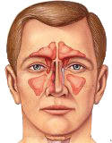
ENT - Ear - Nose - Throat will help you to understand the subject in a better way and also guide you to solve some of common ENT problems.
posted by ENT at
12:21 AM
1 Comments
![]()
![]()
posted by ENT at
12:11 AM
2 Comments
![]()
![]()


posted by ENT at
9:30 PM
0 Comments
![]()
![]()


posted by ENT at
12:21 AM
1 Comments
![]()
![]()


posted by ENT at
6:17 PM
0 Comments
![]()
![]()
posted by ENT at
7:36 PM
0 Comments
![]()
![]()
posted by ENT at
9:09 PM
0 Comments
![]()
![]()
i) Hb, PCV - to see the amount of haemoglobin in blood and packed cell value helpful in acessing the general condition of the patient and to see need for any blood transfusion.
ii) Platelet count - to find any platelet deficiency state.
iii) PTI (prothrombin time index) - access the coagulatory mechanism.
iv) Total and differential leucocytic count - to see for any infection
v) Perepheral blood film - to see any immature cells and deficiency states.
vi) Bleeding and clotting time
2. Radiological investigations -
i) X-ray PNS water's view - to see any sinus pathology, fractures but does not give much information.
ii) CT scan and MRI - In cases of carcinoma and angiofibroma
3. Angiography - To see the vessel which is bleeding.
posted by ENT at
10:47 PM
0 Comments
![]()
![]()
posted by ENT at
11:38 PM
0 Comments
![]()
![]()
posted by ENT at
8:01 PM
0 Comments
![]()
![]()
posted by ENT at
6:52 PM
0 Comments
![]()
![]()
posted by ENT at
11:22 PM
0 Comments
![]()
![]()
2. Traumatic causes -
posted by ENT at
10:17 PM
0 Comments
![]()
![]()


posted by ENT at
12:13 AM
0 Comments
![]()
![]()
posted by ENT at
8:20 PM
0 Comments
![]()
![]()


posted by ENT at
8:45 PM
0 Comments
![]()
![]()
posted by ENT at
12:45 AM
2 Comments
![]()
![]()
The tight sutures act as ball valve. Air gets sucked into subcutaneous tissue but cannot comes out. So it gets collected under skin. If not checked, there can be pneumothorax and pneumomediastinum.
On examination ,on palpation creps can be felt under skin which can also be auscultated.
Treatment - Open the tight sutures. If extensive surgical emphysema is there oh face, neck and shoulders, then small nicks can be given to release air. If there is pnemothorax and pneumomediastinum, they are treated actively.
posted by ENT at
12:51 AM
1 Comments
![]()
![]()
In emergency tracheostomy, vertical skin incision is preferred. After incision, the larynx is fixed with one hand and the incision is deepened not bothering about isthmus of thyroid or anything in the way direct to the trachea and then after identifying it, the trachea is opened,wound dilated and the tracheostomy tube inserted. As soon as the airway is made patent most of the bleeding stops automatically. Rest of the bleeders are secured and skin wound is stitched with silk.
posted by ENT at
11:23 PM
0 Comments
![]()
![]()
posted by ENT at
12:27 AM
0 Comments
![]()
![]()
1.Obstruction of the tracheostomy tube due to -
a) any clot in the tube
b) Any mucus plug in the tube
c) impingement on the posterior tracheal wall
d) partial displacement into the mediastinum or in a false passage
2. Wound infection
3. Tracheal injury and granulation tissue formation.
4. Fatal haemorrhage due to injury to inomanate artery due to continuous irritation and injury causing a tracheo-inominate fistula leading due massive haemorrhage and death.
5. Tracheo-oesophageal fistula.
6. Scar formation and tracheal tug.
7.Difficult decanulation.
posted by ENT at
12:05 AM
0 Comments
![]()
![]()
posted by ENT at
11:49 AM
0 Comments
![]()
![]()

Subscribe to
Posts [Atom]